INTRODUCTION
Dry eyes can result in a variety of ocular discomfort,reducing working and living comfort. According to an epidemiological survey in recent years, the prevalence of dry eye was greater than 10% with a rising trend[1]. Decrease of testosterone levels has been regarded as one of the main reasons for dry eye disease, and often causes dry eye in postmenopausal women and older men[2-3]. If not treated, dry eye will further develop into severe corneal ulcer, corneal perforation, impairing vision and eventually causing loss of vision[4-5]. Before serious symptoms occur, it is important to look for a way to treat and prevent the progression of the dry eye. Clinical treatment of hormone deficiency dry eye is usually the use of hormone replacement treatment, but the efficacy still needs to be further confirmed.
Mistletoe belongs to the loranthaceae family, and tastes bitter.It has been used to treat rheumatism, and has protective effects on the liver, kidney, bone and tocolysis, as described in the pharmacopoeia. Mistletoe mainly contains flavonoids[6], and plays a role in the interaction with sex hormone receptors. Its chemical structure is similar to androgen, and may have similar function to androgen[7]. Unlike androgen, it does not cause complications such as virilization and acne. It can also shorten the duration of symptoms effectively[8]. Mistletoe has a lot of applications. Some studies showed that it has the functions of resisting tumor, lowering blood glucose, regulating blood lipid and immune response[9-11]. In recent years, there are more and more studies on the application of mistletoe in the treatment of eye diseases. Munteanu et al[12] showed that garlic and mistletoe extracts could play a positive role in the treatment of choroidal melanoma.
In a prospective, randomized and controlled study, the presented study treated climacteric female dry eye patients with the mistletoe combined with carboxymethyl cellulose eye drops and compared with normal saline eye drops treatment.At present, there is no report of mistletoe combined with carboxymethyl cellulose eye drops used in the treatment of dry eye in postmenopausal women. This paper aims to introduce such new eye drops to the clinical treatment of climacteric xeroma patients and to explore the safety and effectiveness, in order to better generalize its application.
SUBJECTS AND METHODS
Experimental Subjects Sixty climacteric women patients diagnosed with dry eye were recruited from the Department of Ophthalmology, the First Affiliated Hospital of Nanchang University from October 2014 to October 2015.
The menopausal women recruitment criterions were: 1) female dry eye patients aged 50 to 56y (average age 53.46±3.31y)with typical symptoms of menopausal women: hyperhidrosis and hot flashes or accompanied with these features, increased irritability, palpitation, psychological problem, bad memory and backaches etc. and the disruption of the menstrual cycle;2) endocrine hormone test: low E2 levels and the high levels of FSH and LH; 3) No cardiovascular disease, mental illness and thyroid malfunction etc.
Standard diagnostic conditions were used to determine the dry eye[13]: 1) chronic symptoms such as visual tiredness, dry and unsmooth sensation, foreign body sensation, burning sensation,photophobia, pain, red eye, and various ocular discomforts;2) tear film break-up time (BUT): positive <10s; 3) schirmer I test (SIT): SIT values ≤10 mm/5min; 4) low tear lactoferrin concentration or high osmotic pressure (reinforced diagnosis);5) ocular surface damage (reinforced diagnosis) as located with fluorescein staining (FL).
The study was conducted in accordance with the principles of the Declaration of Helsinki. The study protocol has been approved by the Institutional Review Board. The experiment procedure was fully explained to each patient, and consent was obtained according to the Ethical Committee of the First Affiliated Hospital of Nanchang University with signatures of these patients and the dates marked.
Preparation of Mistletoe Extract Precisely weighted 1 gram of mistletoe powder was put in a conical flask, and 30 mL hydrochloric acid: ethanol solution (1:10) was added. The mixture was weighted and sonicated for 1.5h, then cooled and weighted again. Lost weight was complemented with hydrochloric acid: ethanol solution. The solution was then shaken and filtered. 15 mL filtrate was concentrated to about 5 mL, added with 10 mL water, and then extracted with chloroform 5 times (20 mL, 20 mL, 20 mL, 15 mL and 15 mL).The extract was pooled and evaporated. The residue was dissolved with 5 mL chloroform, and added into the alumina column (diameter 0.9 cm, neutral alumina 5 g, 100-200). The column was washed with 30 mL chloroform: methanol (80:1),and eluted with 50 mL chloroform: methanol (20:1). The elution was collected, evaporated, dissolved in pure ethanol and diluted to 5 mL in a volumetric flask. The solution was the mistletoe extract.
Preparation of Mistletoe Extract Eye Drops The mistletoe extract was diluted in distilled water in 0.1:1 ratio, added in carboxymethyl cellulose with final concentration of 1.5%,and buffered with NaHCO3 and KCL with final concentration less than 0.1%. The pH value, osmotic pressure, specific gravity, surface tension, viscosity and refractive index were measured and adjusted to the following conditions: pH value:7.8; osmotic pressure: 311-350 mOsm; specific gravity:about 1; surface tension: 40-50 dyn/cm2; viscosity: slightly higher than water; refractive index: standard 1.336. Finally,the preservative benzyl bromide was added with controlled concentration of 0.005%.
Clinical Experiment Design Our patients were prospectively randomized in a single-blind, parallel controlled clinical trial.Sixty patients (60 eyes) were randomly divided into two groups with 30 subjects each group (30 eyes). Group A (n=30)received the mistletoe combined with carboxymethyl cellulose eye drops treatment, whereas group B were treated with saline eye drops (n=30). Both groups were dosed 4 times a day for 2mo. The Ocular Surface Disease Index (OSDI), the tear film test with 4 terms, tear proteins were analyzed before the treatment and 7d, 1 and 2mo after the treatment, respectively.And analysis of variance and sample mean or median difference was performed, and the differences were compared with corneal confocal microscopy.
Subjective Symptom Score of the Ocular Surface OSDI questionnaire survey is a well-established method in assessing ocular surface diseases[14]. The patients were asked whether they had discomfort from dryness, abnormal sensation, or asthenia: 0 points represented asymptomatic, 0.5 points represented occasional symptoms, 1 point represented intermittent mild symptoms, and 2 points represented sustained obvious symptoms. To assess symptoms, patients also completed the Chinese version of the OSDI questionnaire[13-14].This 12-item, self-administered questionnaire assessed ocular symptoms (photophobia; gritty sensation; eye pain; blurred vision; poor vision; difficulty in reading, driving at night,working with a computer or automatic teller machine, or watching television; discomfort under windy conditions, in places with low humidity, or in air-conditioned areas) duringthe 2- to 4-week period before the examination[15-16]. OSDI was calculated as the sum of the scores of all questions/number of answers ×25.
Table 1 Baseline characteristics of the participants Mean±SD (range)

Tear Film Test with Four Terms The tear film test including BUT, FL, SIT and the height of the tear meniscus (HTM)was performed as described previously[17]. Tear stability was assessed by performing a BUT evaluation, which measures the time interval between a blink and the appearance of a break in the tear film after the fluorescein staining. flwas measured with an ocular test strip to observe patients’ corneal conditions.In the SIT, a thin strip of filter paper (35 mm×5 mm) was used to quantify tear production over a period of 5min. The strip was placed at the junction of the middle and lateral thirds of the lower eyelid. The patient was instructed to look forward and blink normally during the test. And SIT value <5 mm/5min shows the symptoms of dry eye. The height of the tear meniscus in the central lower eyelid was measured by a ruler under a slit-lamp microscope[18].
Tear Protein Measurements Tear protein levels were measured as described previously[19-20]. All tear samples were collected from 9:00 a.m. to 11:00 a.m. The total tear protein concentration was measured by the Brandford method using bovine serum albumin as the standard. Lactoferrin concentration was measured by radioimmunoassay. Lysozyme concentration was measured with a turbidimeter.
Corneal Confocal Microscopy Corneal confocal microscopy was performed as described[21]. After ocular surface anesthesia,the patient’s eyes were fixed staring straight ahead under the microscope. The examiner slowly pushed the objective forward to focus on the corneal epithelial cells. Full-tomography was obtained from the central cornea. Valuable images and videos were selected and saved, and the density of the corneal epithelial basal cells and inflammatory cells was counted.
Statistical Analysis All calculations and statistical analyses were performed using the 19.0 software package for Windows(SPSS Inc. Chicago, IL, USA). All values were presented as mean±standard deviation (SD). ANOVA was used for all indexes in subjective symptoms before and after treatment comparisons; Dunnetts-test was applied for multiple comparisons. Differences between two groups in age, spherical equivalent refractive error, interval between onset of symptoms and treatment and duration time were performed using the paired t-test. A value of P<0.05 was considered statistically significant.
RESULTS
The Clinical Outcomes In this study, no patients were isolated from the study. There were no statistical differences between two groups in age, BMI, spherical equivalent refractive errors, interval between onset of symptoms and treatment and duration time (P>0.05). The detailed information is presented in Table 1.
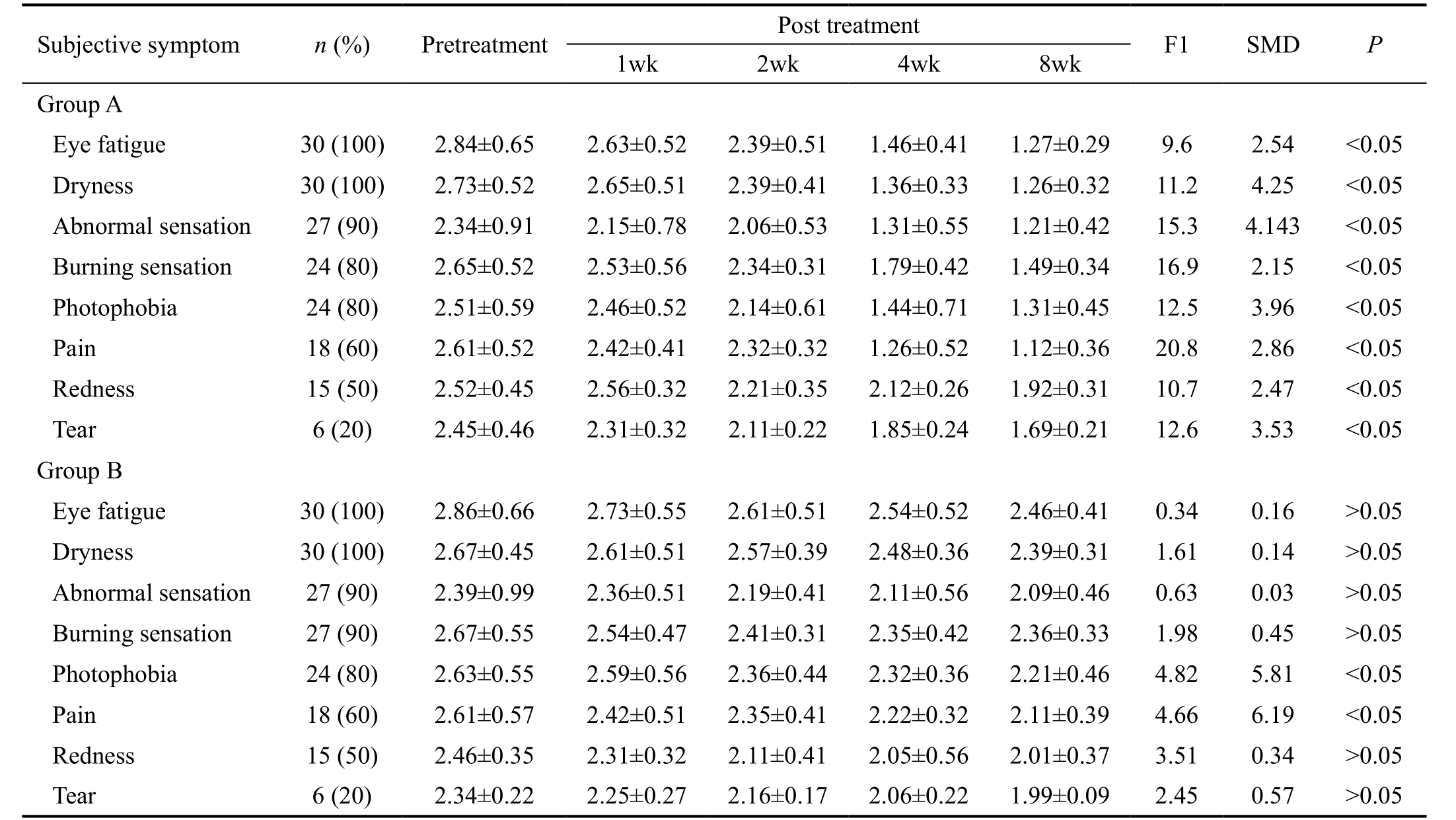
Subjective Symptom of the Ocular Surface No significant differences in the subjective symptom of the ocular surface between two groups were observed before the treatment(P>0.05). At 8wk after treatment, the subjective symptoms of dry eye in group A were improved (P<0.05), while the control group didn’t show significant changes (except photophobia and pain) at 8wk after treatment (P>0.05). The detailed information is presented in Table 2.
The disease symptoms indices were improved in group A but not in group B (P>0.05), according to the results of the OSDI questionnaire. The mean total OSDI scores were presented in Figure 1. The results of OSDI questionnaire showed an improvement of the ocular surface symptoms in the group treated with mistletoe combined with carboxymethyl cellulose eye drops but not with normal saline eye drops (P<0.05) at 8wk after treatment.
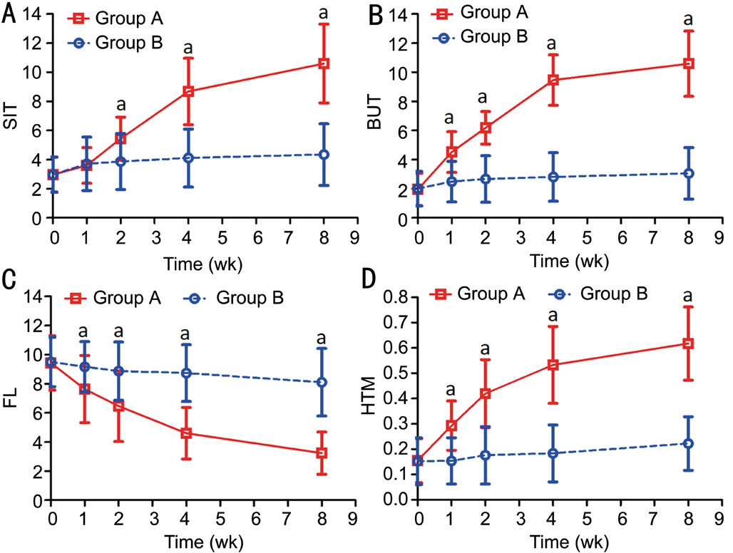
Tear Film Test The results of tear film tests in the two groups before treatment and at 1, 2, 4, and 8wk after treatment were summarized in Figure 2. There were no statistically differences in SIT, BUT, fland HTM between two groups before treatment (P>0.05). The flof group A decreased at 1, 2, 4 and 8wk after treatment (all P<0.05). On the contrary, the BUT,SIT, HTM and flin group B did not change markedly at 8wk after treatment, compared with those of pretreatment (P>0.05)(Figure 2). The result in BUT and HTM of group A was improved at 2, 4 and 8wk after treatment (P<0.05), while there was no significant change at 1wk after treatment compared with those of pretreatment (P>0.05) (Figure 2A).
Tear Proteins Changes of tear proteins in the two groups at pretreatment day and 1, 2, 4 and 8wk after treatment were presented in Figure 3. Before the treatment, there were nostatistically significant differences in the total tear proteins,lactoferrin, lysozyme and lipocalin between two groups(P>0.05). However, at 8wk after the treatment, the tear proteins, lactoferrin, lysozyme as well as lipocalin in the group A showed significantly increase compared with those of group B (P<0.05). The total tear proteins, lactoferrin and lysozyme of group A showed an increase since 4wk after treatment(P<0.05) (Figures 3A, 3B and 3C). And the lipocalin in group A but not group B was significantly improved at 8wk (P<0.05;Figure 3D). On the contrary, there were no remarkably changes at 1, 2 and 4wk between the two groups (P>0.05;Figure 3D).
Table 2 The symptoms of ocular surface at baseline and after treatment

α=0.05/4=0.0125. F1: MD of the symptom of ocular surface pretreatment; SMD: MD of the symptom of ocular surface at 8wk post treatment.

Figure 1 The mean total OSDI scores of eyes on two groups A:The mean total OSDI scores of eyes with postmenopausal patients treated with mistletoe combined with carboxymethyl cellulose eye drops and normal saline eye drops in the treatment time course. The results showed the symptoms of ocular surface were improved in patients treated with mistletoe combined with hyaluronic acid eye drops but not with normal saline eye drops (P<0.05); B: The mean ocular symptoms OSDI score between two groups at 8wk after treatment (P<0.05). aP<0.05 vs group A.

Figure 2 The dynamic change of SIT, BUT, fland HTM before and after treatment There was no significant difference in group B until 8wk treatment (P>0.05). However, the BUT, HTM in group A was remarkably greater than that of group B after 8wk treatment(P<0.05, B, D). The SIT values of group A were greater than that of group B at 2, 4 and 8wk, although there was no significant difference at 1wk after treatment (P<0.05) A: The flof group A was reduced compared with Group B at 1, 2, 4 and 8wk after treatment either(P<0.05); C: n=30 for both groups. aP<0.05 vs group A.

Figure 3 Changes of total tear proteins, lactoferrin, lysozyme and lipocalin before and after the treatment The figure showed that the total tear proteins, lactoferrin, lysozyme of group A were greater than that of group B at 4 and 8wk, while there were no significant differences at 1 and 2wk after treatment (P<0.05) (A, B, C). There was a significant improvement of the lipocalin in group A compared with that of group B at 8wk (P<0.05) (D), but there were no significant changes at 1, 2 and 4wk between the two groups (P>0.05) (D). The sample size was 30 cases for both group A and group B throughout our study. aP<0.05 vs group A.

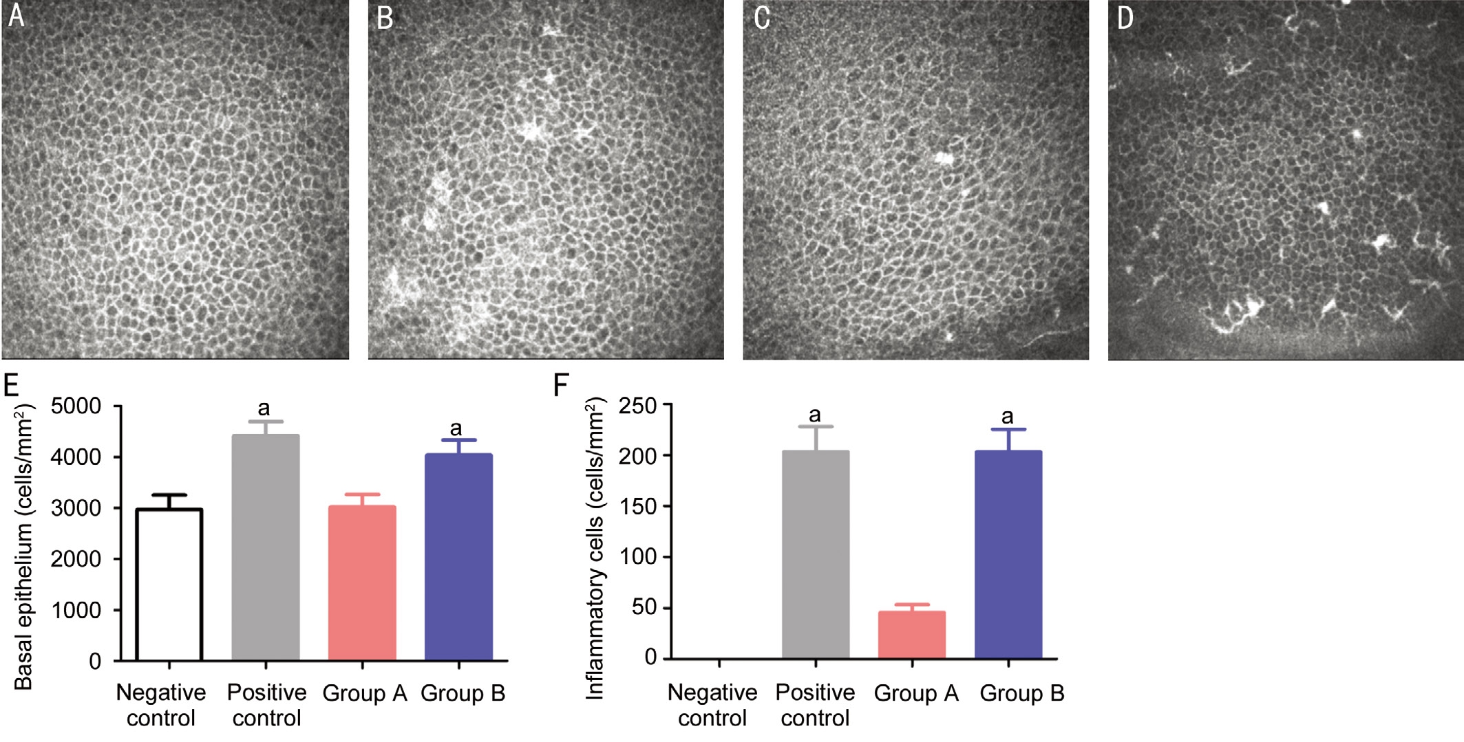
Figure 4 The morphology of female corneal epithelial basal layer after the treatment of mistletoe combined with carboxymethyl cellulose eye drops and normal saline eye drops A: Normal females; B: Postmenopause females with dry eye; C: Postmenopause females treated with mistletoe combined with carboxymethyl cellulose eye drops; D: Postmenopause females treated with normal saline eye drops; E: The mean corneal basal cell densities in four groups. F: The mean inflammatory cell densities in four groups. Data were mean±SD of values from all eyes per group. aP<0.05 vs group A.
Corneal Confocal Microscopy Under the corneal confocal microscopy, the normal female’s corneal epithelial basal cells were dark cells with distinct boundaries (Figure 4A). However,the corneal epithelial basal cells of postmenopausal patients with dry eye were infiltrated with bright contractible atrophied inflammatory cells (Figure 4B). At 2mo after treatment, group A showed less inflammatory cells infiltrated in the epithelial basal layer, compared with group B. Consistently, the density of the corneal epithelial basal cells was decreased slightly compared with that of normal female but much lower than in group B (Figures 4C and 4D). The average amount of corneal epithelium basal cells and inflammatory cells in group A were 3174±379 and 38±25 cells/mm2, while the density of group B of epithelial basal cells and inflammatory cells were 4309±612 and 158±61 cells/mm2, respectively. There were statistically significant differences between two groups (P<0.05; Figure 4E and 4F), and there were no obvious differences between group B and the postmenopausal females regarding corneal epithelial basal cells density and the number of inflammatory cells (P>0.05).
The normal female’s epithelial nerve plexus showed normal course and distribution (Figure 5A). In contrast, the epithelial nerve plexus in postmenopausal females were curving and abnormal (Figure 5B). After two months of treatment, the epithelial nerve plexus in group A (treated with mistletoe combined with carboxymethyl cellulose eye drops) were almost comparable to normal condition (Figure 5C). In contrast, group B (treated with normal saline eye drops)patients still had curving and abnormal epithelial nerve plexus(Figure 5D). In addition, postmenopausal patients with dry eye had more shiny cells in the corneal stroma (Figure 5F)compared to normal female (Figure 5E). After two months of treatment, group A (treated with mistletoe combined with carboxymethyl cellulose eye drops) showed decreased number of shiny cells, and was comparable to normal condition (Figure 5G).In contrast, group B (treated with normal saline eye drops) still maintained many shiny cells in the corneal stroma (Figure 5H).

Figure 5 Representative in vivo confocal images of the epithelial nerve plexus and the corneal stromal cells in different groups A: Normal females; B: Postmenopause females of dry eye; C: Postmenopause females of dry eye treated with mistletoe combined with carboxymethyl cellulose eye drops; D: Postmenopause females of dry eye treated with normal saline eye drops; E: Normal females; F: Postmenopause females of dry eye; G: Postmenopause females of dry eye treated with mistletoe combined with carboxymethyl cellulose eye drops; H: Postmenopause females of dry eye treated with normal saline eye drops.
DISCUSSION
Dry eye is one of the most common chronic ocular surface diseases, characterized by high incidence and multi-factor effects, and has been identified as a worldwide public health problem[22-23]. Dry eye is also known as the dry conjunctivitis,which refers to the falling of tear film stability by the abnormity of tears quality, quantity or dynamics, leading to the damage of the ocular surface structure. Dry eye can be caused by many factors including infection, inflammation, trauma,abnormal eye anatomical structure and high tear osmotic pressure, etc[24]. Without reasonable treatment, it can even lead to blindness.
The dry eye diagnostic classification standard has yet been unified. In 1995, an American dry eye research group classified dry eye into insufficient tear production type and strong evaporation type[21]. According to the etiology, dry eye can also be classified as aqueous tear deficiency, mucin deficiency, lipid tear deficiency and abnormal distribution of tear dynamics[25].
It is helpful to clarify the type of dry eye for specific treatment to achieve better efficacy. At present, dry eye is mainly treated with local symptomatic agents, including artificial tears, glucocorticoid, immunosuppressive eye drops and lacrimal duct embolization[26]. For moderate to severe dry eye patients, the use of autologous isolated submandibular gland transplantation surgery may be possible options[27]. If using artificial tears, attention should be paid to avoid antibiotics and effects of preserving agent contained within the drug, to prevent adverse effects in the diagonal conjunctiva[28].
Dry eye concurs with age in menopausal or postmenopausal women, and the ratio is 9:1 of men and women[29]. But in recent years, with the wide usage of electronic devices and the popularity of refractive surgery, the incidence of dry eye is also significantly increased in the young people[30-31]. For menopausal women, the drop of androgen levels is the main reason for dry eye. Androgen can interact with a variety of hormones, affecting the endocrine process, and it is important to keep androgens in optimal biological level for the function of tear secretion[32]. Currently, androgen replacement treatment is the main method for the treatment of this type of dry eye,but long-term usage of androgen can cause a lot of serious side effects, such as acne, erythrocytosis, male prostate cancer,breast development, virilization etc., which can greatly affect the patient’s life quality[33]. It is imperative to find alternative androgen replacement drugs.
Decreased androgen levels cause the reduction of the secretion of meibomian gland and lacrimal gland. Study found that androgen could prevent apoptosis, necrosis and lymphocytic infiltration of lacrimal gland[34]. Other studies have shown that reduced androgen levels may act through the NF-κB activation to induce the expression of NOS2 gene, and produce large amounts of NO, which damages the ocular surface epithelium,leading to the occurrence and development of dry eye[3]. In some postmenopausal dry eye female patients, androgen levels are reduced, and reduction of secretion from the ovarian causes declined secretion activity of meibomian glands and Zeis gland, reducing the tear film lipid composition and increasing tear evaporation, leading to the elevated incidence of lipid deficiency in menopausal or postmenopausal women[35-36]. It has been proved that the eye is the target organ of sex hormone.Androgen, estrogen, progesterone and prolactin receptor exists widely in human, rabbit and rat lacrimal, meibomian gland,cornea and other ocular surface tissues[37].
In recent years, some research suggests that Buddleja officinalis and Bidens bipinnata L. could be used to not only treat dry eye in a model of androgen decline in female/male rabbit[38], but also obviously reduce the signs and symptoms of moderate and severe dry eye in postmenopausal women, which shows a great clinical value. The flavonoids are effective because of the similar structure to androgen[39]. However, Buddleja officinalis is sweet, which is not suitable for people with diabetes,whereas Bidens bipinnata L. is bitter and has poor patients compliance.
Mistletoe as a long-used traditional Chinese medicine, has been used for the treatment of rheumatism, soreness and weakness of waist and knees, and threatened abortion. It has also been used in folk medicine for the treatment of liver cancer, squamous cell carcinoma and other cancers[40-42].Modern medical research found mistletoe has the effects of anti-arrhythmia, anti-platelet aggregation, anti-thrombosis,increasing coronary blood flow, improving the coronary circulation, enhancing myocardial contractility, reducing myocardial oxygen consumption, prevention and treatment of myocardial infarction, and reducing blood pressure etc[43-44]. It can be regarded as a heart protection medicine in the prevention and treatment of coronary heart disease. In recent years,studies of mistletoe emerge rapidly. Discovery of new effects and new applications, especially obvious anti-tumor effects,provides prospect applications for mistletoe. Demand for mistletoe is also growing[45]. Researchers have isolated 36 flavonoids from mistletoe, including 3’-rhamnazin, 3’-methyl-3-glucose glycosides, mistletoe new glycoside I, II, III, IV,V, VI, VII, 3’-methyl eriodictyol etc[46]. This study showed that the mistletoe combined with carboxymethyl cellulose eye drops could significantly improve the symptoms of dry eye. OSDI questionnaire survey results showed that after two months of treatment, symptoms of eye fatigue, dryness,abnormal sensation, burning sensation, photophobia, pain, and redness etc. were significantly improved in the treatment of mistletoe combined with carboxymethyl cellulose eye drops,possibly due to the inhibition of VEGFR2 signaling pathway.Rhamnazin, the major component of mistletoe, has the effects of anti-angiogenesis, anti-inflammatory and anti-tumor.Because dry eye itself is a kind of inflammation of the eye,mistletoe combined with carboxymethyl cellulose eye drops might also have the anti-inflammatory role in the dry eye[47].Mistletoe has bacteriostatic effects for various kinds of ocular surface microorganisms, and it also contains a lot of choline,which causes tears secretion, and prevents further deterioration of the dry eye. Deeni and Sadiq[48] found that mistletoe had broad-spectrum antibacterial activity, even for some drug resistant bacteria and fungi. In the meanwhile, the lectin and phospholipid in mistletoe can promote the immune system,and improve the antibacterial ability.
Our early study showed that mistletoe eye drops could be very effective in preventing dry eye in castrated rabbits and peri menopausal female rabbits[49-51]. On the basis of that, this study showed that mistletoe combined with carboxymethyl cellulose eye drops can effectively treat dry eye in menopausal women induced by decline of sex hormone.
Our study found that group A patients treated with mistletoe combined with carboxymethyl cellulose eye drops for 2mo showed improved SIT, BUT, fland HTM, possibly because flavonoids compounds in mistletoe extract played the role similar as sex hormone, through the interaction with ocular surface tissue hormone receptors and promoting the secretion of eyelid glands. Mistletoe extracts eye drops contain high content of flavonoid compounds, so it might indirectly or directly produce the estrogenic activity. In addition, the alkaloids in the mistletoe extract have certain antibacterial function, and the toxin can improve immune function, so these might also play certain roles in the mitigation of the dry eye syndrome. Two months after treatment in group A, total tear proteins, lactoferrin, lysozyme and amylase activity were improved, further suggesting that mistletoe combined with carboxymethyl cellulose eye drops have certain therapeutic effects.
Confocal microscopy can effectively evaluate the dynamic changes of various cells in the cornea. In this study, we compared the corneal epithelial basal cells, the corneal epithelium and the anterior stromal cells of the corneal epithelium by corneal confocal microscopy. Normal corneal epithelial basal cells have light dark cell bodies, with a narrow cell border, and the epithelial nerves are straight with less corneal stromal cells. Results showed that in group A with mistletoe combined with carboxymethyl cellulose eye drops treatment for 2mo, corneal epithelial basal layer cells were reduced in size, inflammatory cell infiltration was decreased, epithelial basal cells and inflammatory cell density declined, and corneal subepithelial nerve was straight and subcutaneous nerve quantity was slightly reduced compared to normal women. Corneal stromal cells were decreased,and gradually tended to be normal. In comparison, group B showed a tendency of deterioration, suggesting that in mistletoe combined with carboxymethyl cellulose eye drops treated corneal cells, nerve degeneration was significantly less than that of saline eye drops treatment. As mistletoe belongs to natural traditional Chinese medicine, it can improve ocular symptoms without obvious ocular and systemic side effects in the treatment of dry eye.
In summary, mistletoe combined with carboxymethyl cellulose eye drops can quickly and markedly improve the symptoms of dry eye and maintain tear protein component, effectively relieve the signs and symptoms of dry eyes in menopausal women. The method of the treatment of dry eye is convenient and simple, and has good clinical application values. In terms of serious dry eye, further investigation will be needed to determine whether mistletoe treatment is still effective and whether symptoms will relapse after discontinuation of the drug, and the side effects of long-term medication.
ACKNOWLEDGEMENTS
Foundations: Supported by the National Natural Science Foundation of China (No.81460092, No.81660158 and No.81400372); Natural Science Key Project of Jiangxi Province (No.20161ACB21017); Youth Science Foundation of Jiangxi Province (No.20151BAB215016); Technology and Science Foundation of Jiangxi Province (No.20151BBG70223)Conflicts of Interest: Jiang N, None; Ye LH, None; Ye L,None; Yu J, None; Yang QC, None; Yuan Q, None; Zhu PW,None; Shao Y, None.
REFERENCES
1 Sahai A, Malik P. Dry eye: prevalence and attributable risk factors in a hospital-based population. Indian J Ophthalmol 2005;53(2):87-91.
2 Gipson IK, Spurr-Michaud SJ, Senchyna M, Ritter R 3rd, Schaumberg D. Comparison of mucin levels at the ocular surface of postmenopausal women with and without a history of dry eye. Cornea 2011;30(12):1346-1352.
3 Azcarate PM, Venincasa VD, Feuer W, Stanczyk F, Schally AV, Galor A. Androgen deficiency and dry eye syndrome in the aging male. Invest Ophthalmol Vis Sci 2014;55(8):5046-5053.
4 Miljanović B, Dana R, Sullivan DA, Schaumberg DA. Impact of dry eye syndrome on vision-related quality of life. Am J Ophthalmol 2007;143(3):409-415.
5 Liu Z, Pflugfelder SC. Corneal thickness is reduced in dry eye. Cornea 1999;18(4):403-407.
6 Lobo R, Sodde V, Dashora N, Gupta N, Prabhu K. Quantification of flavonoid and phenol content from Macrosolen parasiticus (L.) Danser.Nat Prod Plant Resour 2011;1(4):96-99.
7 Ofem OE, Antai AB, Essien NM, Oka VO. Enhancement of some sex hormones concentrations by consumption of leaves extract of Viscum album (mistletoe) in rats. Asian Journal of Medical Sciences 2014;5(3):14-16.
8 Korman DB. Mistletoe lectins-antitumor effect and mechanism of action. Vopr Onkol 2011;57(6):689-698.
9 Eisenbraun J, Scheer R, Kröz M, Schad F, Huber R. Quality of life in breast cancer patients during chemotherapy and concurrent therapy with a mistletoe extract. Phytomedicine 2011;18(2-3):151-157.
10 Abdel-Sattar E, Harraz FM, Ghareib SA, Elberry AA, Gabr S,Suliaman MI. Antihyperglycaemic and hypolipidaemic effects of the methanolic extract of Caralluma tuberculata in streptozotocin-induced diabetic rats. Nat Prod Res 2011;25(12):1171-1179.
11 Braun JM, Ko HL, Schierholz JM, Weir D, Blackwell CC, Beuth J.Application of standardized mistletoe extracts augment immune response and down regulates metastatic organ colonization in murine models.Cancer Lett 2001;170(1):25-31.
12 Munteanu MF, Ardelean A, Borcan F, Trifunschi SI, Gligor R,Ardelean SA, Coricovac D, Pinzaru I, Andrica F, Borcan LC. Mistletoe and garlic extracts as polyurethane carriers - a possible remedy for choroidal melanoma. Curr Drug Deliv 2017:14(999).
13 Zhang X, Zhao L, Deng S, Sun X, Wang N. Dry eye syndrome in patients with diabetes mellitus: prevalence, etiology, and clinical characteristics. J Ophthalmol 2016;2016(2):1-7.
14 Schiffman RM, Christianson MD, Jacobsen G, Hirsch JD, Reis BL. Reliability and validity of the Ocular Surface Disease Index. Arch Ophthalmol 2000;118(5):615-621.
15 Walt JG, Ravelo AL, Lee JT, Lee L. Assessing the functional impact and severity of ocular surface disease using the Ocular Surface Disease Index (OSDI©). The Ocular Surface 2005;3(12):S124-S124.
16 Miller KL, Walt JG, Mink DR, Satram-Hoang S, Wilson SE, Perry HD, Asbell PA, Pflugfelder SC. Minimal clinically important difference for the ocular surface disease index. Arch Ophthalmol 2010;128(1):94-101.
17 de Souza GA, Godoy Lyri MF, Matthias M. Identification of 491 proteins in the tear fluid proteome reveals a large number of proteases and protease inhibitors. Genome biology 2006;7(8):R72.
18 Sakane Y, Yamaguchi M, Yokoi N, Uchino M, Dogru M, Oishi T,Ohashi Y, Ohashi Y. Development and validation of the Dry Eye-Related Quality-of-Life Score questionnaire. JAMA Ophthalmol 2013;131(10):1331-1338.
19 Srinivasan S, Joyce E, Boone A, Simpson T, Jones L, Senchyna M.Tear lipocalin and lysozyme concentrations in postmenopausal women.Ophthalmic Physiol Opt 2010;30(3):257-266.
20 Chen W, Li Z, Hu J, Zhang Z, Chen L, Chen Y, Liu Z. Corneal alternations induced by topical application of benzalkonium chloride in rabbit. PLoS One 2011;6(10):e26103.
21 Galor A, Levitt RC, Felix ER, Martin ER, Sarantopoulos CD.Neuropathic ocular pain: an important yet underevaluated feature of dry eye. Eye (Lond) 2015;29(3):301-312.
22 Bakkar MM, Shihadeh WA, Haddad MF, Khader YS. Epidemiology of symptoms of dry eye disease (DED) in Jordan: A cross-sectional nonclinical population-based study. Cont Lens Anterior Eye 2016;39(3):197-202.
23 Osae AE, Gehlsen U, Horstmann J, Siebelmann S, Stern ME, Kumah DB, Steven P. Epidemiology of dry eye disease in Africa: the sparse information, gaps and opportunities. Ocul Surf 2017;15(2):159-168.
24 Jin KW, Ro JW, Shin YJ, Hyon JY, Wee WR, Park SG. Correlation of vitamin D levels with tear film stability and secretion in patients with dry eye syndrome. Acta Ophthalmol 2017;95(3):e230-e235.
25 The definition and classification of dry eye disease: report of the definition and classification subcommittee of the international dry eye workshop (2007). Ocul Surf 2007;5(2):75-92.
26 Alves M, Fonseca EC, Alves MF, Malki LT, Arruda GV, Reinach PS,Rocha EM. Dry eye disease treatment: a systematic review of published trials and a critical appraisal of therapeutic strategies. The Ocular Surface 2013;11(3):181-192.
27 Geerling G, Sieg P, Bastian GO, Laqua H. Transplantation of the autologous submandibular gland for most severe cases of keratoconjunctivitis sicca. Ophthalmology 1998;105(2):327-335.
28 Dutescu RM, Panfil C, Schrage N. Comparison of the effects of various lubricant eye drops on the in vitro rabbit corneal healing and toxicity. Exp Toxicol Pathol 2017;69(3):123-129.
29 Beckman KA, Luchs J, Milner MS, Ambrus JL Jr. The potential role for early biomarker testing as part of a modern, multidisciplinary approach to sjögren's syndrome diagnosis. Adv Ther 2017;34(4):799-812.
30 Molina-Leyva I, Molina-Leyva A, Bueno-Cavanillas A. Efficacy of nutritional supplementation with omega-3 and omega-6 fatty acids in dry eye syndrome: a systematic review of randomized clinical trials. Acta Ophthalmologica 2017.
31 Kandel H, Khadka J, Goggin M, Pesudovs K. A pair of glasses and beyond: impact of refractive error on people’s well-being. Clin Exp Ophthalmol 2017;45(7):677-688.
32 Vehof J, Hysi PG, Hammond CJ. A metabolome-wide study of dry eye disease reveals serum androgens as biomarkers. Ophthalmology 2017;124(4):505-511.
33 Handelsman DJ. Androgen misuse and abuse. Best Pract Res Clin Endocrinol Metab 2011;25(2):377-389.
34 Azzarolo AM, Wood RL, Mircheff AK, Richters A, Olsen E, Berkowitz M, Bachmann M, Huang ZM, Zolfagari R, Warren DW. Androgen influence on lacrimal gland apoptosis, necrosis, and lymphocytic infiltration. Invest Ophthalmol Vis Sci 1999;40(3):592-602.
35 Rocha EM, Wickham LA, da Silveira LA, Krenzer KL, Yu FS, Toda I, Sullivan BD, Sullivan DA. Identification of androgen receptor protein and 5alpha-reductase mRNA in human ocular tissues. Br J Ophthalmol 2000;84(1):76-84.
36 Yu J, Zheng XL, Li YY, Hu PH, Liu RQ, Jiang N, Pei CG, Shao Y. Clinical findings associated with Bidens bipinnata L. eye drops on moderate and severe dry eye in postmenopausal women. Int J Clin Exp Med 2016;9(3):5643-5654.
37 Wickham LA, Gao J, Toda I, Rocha EM, Ono M, Sullivan DA.Identification of androgen, estrogen and progesterone receptor mRNAs in the eye. Acta Ophthalmol Scand 2000;78(2):146-153.
38 Shao Y, Yu Y, Yu J, Pei CG, Gao GP, Tu P. Experimental study on efficiency of Spanishneedles Herb eye drops in treating perimenopausal xerophthalmia in rabbits. Zhongguo zhongyao zazhi 2015;40(6):1151-1155.
39 Shao Y, Yu Y, Huang GD, Tan G, Pei CG, Liu XH. Clinical study on spanishneedles leaves in treatment of middle and severe xerophthalmia of menopausal females. Zhongguo zhongyao zazhi 2012;37(19):2985-2989.
40 Al-Gayyar MM, Ebrahim MA, Shams ME. Measuring serum levels of glycosaminoglycans for prediction and using viscum fraxini-2 for treatment of patients with hepatocellular carcinoma. Journal of Pharmacy Research 2013;7(7):571-575.
41 Werthmann PG, Sträter G, Friesland H, Kienle GS. Durable response of cutaneous squamous cell carcinoma following high-dose peri-lesional injections of Viscum album extracts-a case report. Phytomedicine 2013;20(3-4):324-327.
42 Tröger W, Galun D, Reif M, Schumann A, Stanković N, Milićević M. Viscum album[L.] extract therapy in patients with locally advanced or metastatic pancreatic cancer: a randomised clinical trial on overall survival. Eur J Cancer 2013;49(18):3788-3797.
43 Agrawal M, Nandini D, Sharma V, Chauhan NS. Herbal remedies for treatment of hypertension. Int J Pharm Sci and Res 2010;1(5):1-21.
44 Rysz J, Franczyk B, Banach M, Gluba-Brzozka A. Hypertensioncurrent natural strategies to lower blood pressure. Curr Pharm Des 2017;23(17):2453-2461.
45 Glickman-Simon R, Pettit J. Viscum album (mistletoe) for pancreatic cancer, electromagnetic field therapy for osteoarthritis, homeopathy for multidrug-resistant tuberculosis, vitamin D for depression, acupuncture for insomnia. Explore (NY) 2015;11(3):231-235.
46 Fuchsjäger-Mayrl G, Nepp J, Schneeberger C, Sator M, Dietrich W, Wedrich A, Huber J, Tschugguel W. Identification of estrogen and progesterone receptor mRNA expression in the conjunctiva of premenopausal women. Invest Ophthalmol Vis Sci 2002;43(9):2841-2844.47 Yu Y, Cai W, Pei CG, Shao Y. Rhamnazin, a novel inhibitor of VEGFR2 signaling with potent antiangiogenic activity and antitumor efficacy. Biochem Biophys Res Commun 2015;458(4):913-919.
48 Deeni YY, Sadiq NM. Antimicrobial properties and phytochemical constituents of the leaves of African mistletoe (Tapinanthus dodoneifolius(DC) Danser) (Loranthaceae): an ethnomedicinal plant of Hausaland,Northern Nigeria. J Ethnopharmacol 2002;83(3):235-240.
49 Ye LH, Ye L, Tang LY, Jiang N, Gao GP, Shao Y. Experimental study on efficiency of mistletoe eye drops in treating xerophthalmia of castrated male rabbits. Rec Adv Ophthalmol 2016;36(10):915-918,922.
50 Ye L, Ye LH, Zhang Y, Wei R, Jiang N, Pei CG, Gao GP, Shao Y.Experimental study on mistletoe eye drops in prevention of xerophthalmia of castrated male rabbits. China Journal of Modern Medicine 2017;27(7):20-24.
51 Wang GL, Ye LH, Ye L, Yang QC, Jiang N, Shao Y. Effects of mistletoe eye drops in treating xerophthalmia of perimenopausal female rabbits. Chin Hosp Pharm J 2017;37(9):830-833.