INTRODUCTION
The introduction of intravitreal injections in ophthalmology has revolutionized the treatment of ophthalmic disease.Intravitreal injections have allowed for a high concentration of pharmaceuticals to be instilled at the site of treatment with minimal systemic side effects. Yet, there are numerous local side effects which have yet to be addressed with current day therapeutics. One of the most common side effects of intravitreal therapy is elevated intraocular pressure,both transient and sustained[1-5]. Numerous studies[6-8] have undertaken the task of attempting to decrease the intraocular pressure pre-injection by using anti-hypertensive ocular drops without success. In this case series, the authors investigate the safety and efficacy of a therapeutic anterior chamber paracentesis following an intravitreal injection to decrease the time of elevated intraocular pressure and prevent unwanted side effects related to increased intraocular pressure.
METHODS
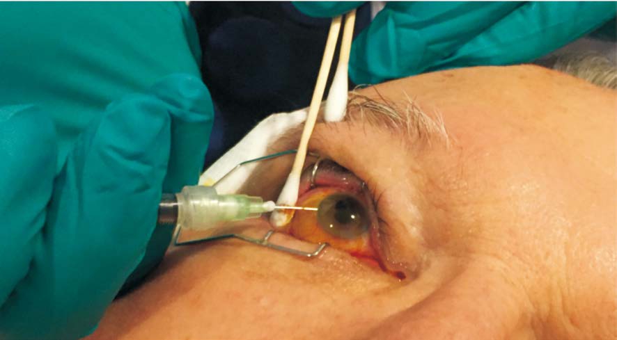
This study is a retrospective analysis approved by the local institutional review board and was conducted in accordance with the Declaration of Helsinki for research in human subjects. The study reviewed the records of all patients that received a therapeutic anterior chamber paracentesis following an intravitreal injection of either bevacizumab or preservativefree triamcinolone acetonide (Figure 1). The patients that were included were those receiving an intravitreal injection with a history of ocular hypertension, primary or secondary open angle glaucoma, or sustained intraocular pressure following a previous injection who presented to a private retina practice over a period of 3y.
RESULTS AND DISCUSSION
Of 1661 anterior chamber paracentesis procedures were performed (Figure 1) on 434 eyes of 395 patients. The average(SD) age was 66.29 (15.75)y. There were 181 (46%) male patients and 214 (54%) female patients. There were a total of 219 (50%) right eyes and 215 (50%) left eyes. Totally 219(13%) of the injections were performed on phakic patients. Of the 395 patients, there was an average of 4.21 injections with a paracentesis per patient.
The average pre-injection intraocular pressure was 22 mm Hg(7.8 mm Hg), and post-injection was 9 mm Hg (9.8 mm Hg).Post injection pressures were measured within the first five minutes following the paracentesis. A median of 210 μL (40 μL)of aqueous was removed during the paracentesis. There were a total of 530 (32%) injections of triamcinolone combined with a paracentesis and 1131 (68%) injections of bevacizumab. Of the 395 patients, 335 had previously received an intravitreal injection. Of 113 (34%) had been injected, at least once, with only bevacizumab, 53 (16%) had been injected, at least once,with only triamcinolone, and 169 (50%) had been injected, at least once, with both bevacizumab and triamcinolone. There were no reported incidences of endophthalmitis, capsular rupture, wound leak, spontaneous cataract formation or any other negative outcome.

Figure 1 Anterior chamber paracentesis of the right eye following an intravitreal injection.
The reasons for intravitreal injection included radiation retinopathy (162, 37%), macular edema (138, 31.8%),neovascular age-related macular degeneration (36, 8.29%),diabetic macular edema (25, 5.8%), retinal vein occlusion (22,5%), proliferative diabetic retinopathy (22, 5%), vitreomacular traction (17, 4%), coats disease (6, 1%), polypoidal choroidal vasculopathy (3, <1%), and sickle cell retinopathy (3, <1%).
The recent revolution of treating ocular pathology with intravitreal injections has changed the face of treating ocular disease. Yet, there has been little done to prevent the most common and unwanted side effect of transient or sustained elevation of intraocular pressure. Transient or sustained intraocular pressure can lead to damage of various ocular structures. There have been a number of cohort studies measuring intraocular pressure following intravitreal antivascular endothelial growth factor injections in patients without glaucoma at different intervals. These studies have shown that the peak intraocular pressure following injection was within the first five minutes, with pressures as high as 87[1-3,5].For this reason, the authors have decided that an immediate anterior chamber paracentesis will alleviate this early peak in pressure as the likely mechanism is an increase in volume inside the globe.
They all showed that the majority of patients’ intraocular pressures returned to the normal range within 30min to one hour and remained at that level for the following week[1-5]. Antivascular endothelial growth factor injections have previously been thought to only cause a transient elevation in intraocular pressure due to a direct intraocular volume increase[1-10],though there have been numerous cases[11-14] showed sustained elevated pressure with intravitreal ranibizumab injections.Intravitreal steroids, on the other hand, have been known to cause both transient elevation as well as sustained elevation of the intraocular pressure. This sustained elevation in intraocular pressure is likely a result of increased outflow resistance[15].This can cause glaucomatous damage by decreasing the ocular perfusion pressure (mean arterial pressure-intraocular pressure)[1,16], as well as retinal vein and/or artery occlusion by direct compression[17-19]. Multiple randomized controlled trials have attempted to prevent this rapid rise in intraocular pressure with no success[6-8].
Performing an anterior chamber paracentesis following injection causes a transient decrease in intraocular pressure.Normal production of aqueous humor can be as much as 3.0 μL/min[20]. At this rate, it should take longer than the hour that it normally takes for the intraocular pressure to return to normal physiologic levels after an anterior chamber paracentesis. Successful performance of this procedure leads to minimal amount of time, less than 5s, of dangerously elevated intraocular pressure. This decreases the likelihood of glaucomatous damage[21] by maintaining physiologic ocular perfusion, and retinal vein and artery occlusion by decreasing the time of increased pressure on the vessels.
There is always the concern of iatrogenic events following any invasive procedure. The likely events include post-operative endophthalmitis, penetration of the crystalline lens in phakic patients, and wound leak. Although there were no cases of any intraoperative or postoperative complications, it is of the utmost importance to follow current day guidelines to prevent a septic work environment and have a physician experienced in intraocular injections performing these procedures to prevent any unwanted side effects of the procedure.
We propose that performing an anterior chamber paracentesis immediately following an intravitreal injection for patients at risk for local side effects such as ocular hypertension,glaucoma, and retinal vein and artery occlusion is a safe and effective procedure. This procedure is not only effective in decreasing the time of elevated intraocular pressure and preventing damage to ocular structures, but it is safe and quick as well. As these patients are already dealing with sight threatening conditions, it is comforting to be able to provide an extra procedure to prevent complications to this sight restoring procedure. Future studies are needed to understand the long term and short term causation and effects of elevated intraocular pressure and determine if a concomitant anterior chamber paracentesis is, in fact, the proper way to prevent intraocular damage from elevated pressures.
ACKNOWLEDGEMENTS
Conflicts of Interest: Bach A, None; Filipowicz A, None;Gold A, None; Latiff A, None; Murray T, None.
REFERENCES
1 Fuest M, Kotliar K, Walter P, Plange N. Monitoring intraocular pressure changes after intravitreal Ranibizumab injection using rebound tonometry.Ophthalmic Physiol Opt 2014;34(4):438-444.
2 Gismondi M, Salati C, Salvetat ML, Zeppieri M, Brusini P. Short-term effect of intravitreal injection of Ranibizumab (Lucentis) on intraocular pressure. J Glaucoma 2009;18(9):658-661.
3 Hollands H, Wong J, Bruen R, Campbell RJ, Sharma S, Gale J.Short-term intraocular pressure changes after intravitreal injection of bevacizumab. Can J Ophthalmol 2007;42(6):807-811.
4 Kiddee W, Montriwet M. Intraocular pressure changes in nonglaucomatous patients receiving intravitreal anti-vascular endothelial growth factor agents. PLoS One 2015;10(9):e0137833.
5 Kim JE, Mantravadi AV, Hur EY, Covert DJ. Short-term intraocular pressure changes immediately after intravitreal injections of anti-vascular endothelial growth factor agents. Am J Ophthalmol 2008;146(6):930-934.
6 Carnota-Mendez P, Mendez-Vazquez C, Otero-Villar J, Saavedra-Pazos JA. Effect of prophylactic medication and influence of vitreous reflux in pressure rise after intravitreal injections of anti-VEGF drugs. Eur J Ophthalmol 2014;24(5):771-777.
7 Frenkel MPC, Haji SA, Frenkel REP. Effect of prophylactic intraocular pressure-lowering medication on intraocular pressure spikes after intravitreal injections. Arch Ophthalmol 2010;128(12):1523-1527.
8 Ozcaliskan S, Ozturk F, Yilmazbas P, Beyazyildiz O. Effect of dorzolamide-timolol fixed combination prophylaxis on intraocular pressure spikes after intravitreal bevacizumab injection. Int J Ophthalmol 2015;8(3):496-500.
9 Lanzl I, Kotliar K. Can anti-VEGF injections cause glaucoma or ocular hypertension? Klin Monbl Augenheilkd 2017;234(2):191-193.
10 Trehan HS, Kaushik J, Rangi A, Parihar AS, Vashisht P, Parihar JKS.Anterior segment changes on ultrasound biomicroscopy after intravitreal anti vascular endothelial growth factor injection. Med J Armed Forces India 2017;73(1):58-64.
11 Reis GM, Grigg J, Chua B, Lee A, Lim R, Higgins R, Martins A,Goldberg I, Clement CI. Incidence of intraocular pressure elevation following intravitreal ranibizumab (lucentis) for age-related macular degeneration. J Curr glaucoma Pract 2017;11(1):3-7.
12 Matsubara H, Miyata R, Kobayashi M, Tsukitome H, Ikesugi K,Kondo M. A case of sustained intraocular pressure elevation after multiple intravitreal injection of ranibizumab and aflibercept for neovascular agerelated macular degeneration. Case Rep Ophthalmol 2016;7(1):230-236.
13 Dedania VS, Bakri SJ. Sustained elevation of intraocular pressure after intravitreal anti-VEGF agents: what is the evidence? Retina 2015;35(5):841-858.
14 Singh RSJ, Kim JE. Ocular hypertension following intravitreal antivascular endothelial growth factor agents. Drugs Aging 2012;29(12):949-956.
15 Kiddee W, Trope GE, Sheng L, Beltran-Aqullo L, Smith M, Strungaru MH, Baath J, Buys YM. Intraocular pressure monitoring post intravitreal steroids: a systematic review. Surv Ophthalmol 2013;58(4):291-310.
16 He Z, Nguyen CTO, Armitage Ja, Vingrys AJ, Bui BV. Blood pressure modifies retinal susceptibility to intraocular pressure elevation. PLoS One 2012;7(2):e31104.
17 Hayreh SS, Zimmerman MB, Beri M, Podhajsky P. Intraocular pressure abnormalities associated with central and hemicentral retinal vein occlusion. Ophthalmology 2004;111(1):133-141.
18 Fieß A, Cal Ö, Kehrein S, Halstenberg S, Frisch I, Steinhorst U.Anterior chamber paracentesis after central retinal artery occlusion: a tenable therapy? BMC Ophthalmol 2014;14(1):28.
19 Natural history and clinical management of central retinal vein occlusion. The Central Vein Occlusion Study Group. Arch Ophthalmol 1997;115(4):486-491.
20 Goel M, Picciani RG, Lee RK, Bhattacharya SK. Aqueous humor dynamics: a review. Open Ophthalmol J 2010;4:52-59.
21 Enders P, Sitnilska V, Altay L, Schaub F, Muether PS, Fauser S.Retinal nerve fiber loss in anti-VEGF therapy for age-related macular degeneration can be decreased by anterior chamber paracentesis.Ophthalmologica 2017;237(2):111-118.